
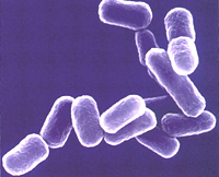
A living photographic “paper” that uses bacteria to produce images has been created by students at The University of Texas at Austin and UCSF. These first-ever bacterial photographs were created using Petri dishes full of genetically engineered E. coli, the students report inNature.
The bacterial camera came about as part of MIT’s intercollegiate Genetically Engineered Machine (iGEM) competition, which encourages students to build simple biological machines. “The goal of the contest was to build bacteria that could do very simple computing,” said Dr. Edward Marcotte, associate professor of biochemistry at The University of Texas at Austin. “This is a great example of the emergent field of synthetic biology – using principles of engineering in biology.”
E. coli are normally found in the dark confines of the human gut and wouldn’t normally sense light, so the students had to engineer the unicellular machines to work as a light sensitive photo-capturing surface. Much as pixels on a computer screen switch between white and black, each bacterium either produced a black pigment or didn’t, depending on whether it was growing in a light or dark place in the dish. The resulting images are a collection of all the bacteria responding to the pattern of light.
The Texas students realized that after optimizing the pigments and growth media, the bacteria could be used to convert light images into biochemical prints. To create the photographs, they used a unique light projector that projects the image onto the dish of bacteria growing in an incubator. But fast shutter speeds aren’t available as exposures take between 12-15 hours (the time it takes for a bacterial population to grow and fill the Petri dish). After exposure, what’s left is a living photograph.
The students are already busy on their next innovation – bacteria that can find and create a line around the edges of an image, a process that requires the bacteria to communicate with each other.


Comments are closed.